
Fayl:PET-image.jpg

Bu prevyuning hajmi: 531 × 600 piksel. Boshqa oʻlchamlari: 1 002 × 1 132 piksel.
Asl fayl (1 002 × 1 132 piksel, fayl hajmi: 134 KB, MIME tipi: image/jpeg)
Qisqa izoh
| Taʼrif |
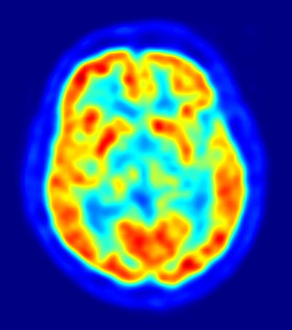
English: This is a transaxial slice of the brain of a 56 year old patient (male) taken with positron emission tomography (PET). The injected dose have been 282 MBq of 18F-FDG and the image was generated from a 20 minutes measurement with an ECAT Exact HR+ PET Scanner. Red areas show more accumulated tracer substance (18F-FDG) and blue areas are regions where low to no tracer have been accumulated.
العربية: صورة مقطعية للدماغ البشري تظهر استهلاك الطاقة. |
||
| Sanasi | |||
| Manba | Oʻzimning ishim | ||
| Muallif | Jens Maus (http://jens-maus.de/) | ||
| Ruxsat (Bu faylning takror foydalanilishi) |
|


Fayl tarixi
Faylning biror paytdagi holatini koʻrish uchun tegishli sana/vaqtga bosingiz.
| Sana/Vaqt | Miniatura | Oʻlchamlari | Foydalanuvchi | Izoh | |
|---|---|---|---|---|---|
| joriy | 01:55, 2026-yil 27-iyul | | 1 002 × 1 132 (134 KB) | wikimediacommons>-sasha- | Reverted to version as of 14:36, 16 March 2010 (UTC) |
Fayllarga ishoratlar
Bu faylga quyidagi 3 sahifalar bogʻlangan:
